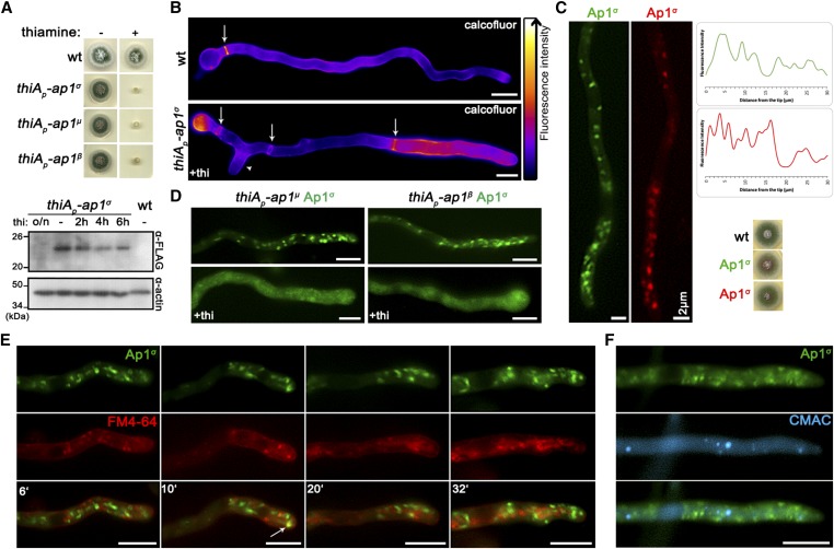
Figure 1.
The AP-1 complex localizes in distinct polarly distributed structures and is essential for growth. (A) Upper panel: Growth of isogenic strains carrying thi-repressible alleles of ap1σ, ap1μ, and ap1β (thiAp-ap1σ, thiAp-ap1μ, and thiAp-ap1β) compared to wild-type (wt) in the absence (−) or presence (+) of thi. Lower panel: Western blot analysis comparing protein levels of FLAG-Ap1σ in the absence (0 hr) or presence of thi, added for 2, 4, 6, or 16 hr (overnight culture, o/n). wt is a standard wild-type strain (untagged ap1σ) which is included as a control for the specificity of the α-FLAG antibody. Equal loading is indicated by actin levels. (B) Microscopic morphology of hyphae in a strain repressed for ap1σ expression (+thi, lower panel) compared to wt (upper panel) stained with calcofluor white. Septal rings and side branches are indicated by arrows and arrowheads. Notice the differences in the calcofluor deposition at the hyphal head, tip, and the sub apical segment (Lookup table [LUT] fire [ImageJ, National Institutes of Health]). (C) Subcellular localization of Ap1σ-GFP and Ap1σ-mRFP in isogenic strains and relative quantitative analysis of fluorescence intensity (right upper panel), highlighting the polar distribution of Ap1σ. Growth tests showing that the tagged versions of Ap1σ are functional (right lower panel). (D) Subcellular localization of Ap1σ-GFP in isogenic strains carrying thi-repressible alleles of ap1μ (left panels) or ap1β (right panels) in the absence (upper panels) or presence of thi (+thi, o/n). Note that repression of expression of either the μ or the β subunit leads to diffuse cytoplasmic fluorescence of Ap1σ. (E) Subcellular localization of Ap1σ-GFP in the presence of FM4-64, which labels dynamically endocytic steps (PM, EEs, late endosomes/vacuoles). Notice that Ap1σ-GFP structures do not colocalize with FM4-64, except a few cases observed in the subapical region (indicated with an arrow at the 10 min picture). (F) Subcellular localization of Ap1σ-GFP in the presence of the vacuolar stain 7-amino-4-chloromethylcoumarin (Blue CMAC). No Ap1σ-GFP/CMAC colocalization is observed. Unless otherwise stated, Bar, 5 μm. Except (C) where the hyphal apex is at the lower side, in all other cases the hyphal apex is at the right side of the image series.

