Figure 3.
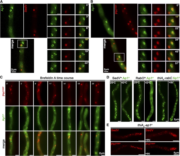
AP-1 associates transiently with the late-Golgi. (A and B) Subcellular localization of Ap1σ-GFP relative to cis- (SedV-mCherry) and trans-Golgi (PHOSBP-mRFP) markers. Notice that Ap1σ colocalizes significantly with the trans-Golgi marker PHOSBP (n = 7; PCC = 0.66, P < 0.0001), but not with the cis-Golgi marker SedV (n = 5; PCC = 0.35, P < 0.01). This colocalization is dynamic and transient, as shown in selected time-lapse images on the right panels (for relevant videos, see also File S1 and File S2). (C) Subcellular localization of Ap1σ and PHOSBP in the presence of the inhibitor Brefeldin A, showing that a fraction of Brefeldin bodies (i.e., collapsed Golgi membranes) includes both markers, further supporting a transient AP-1/late Golgi association. (D) Subcellular localization of Ap1σ in SedVts or RabOts thermosensitive mutants or a strain carrying a repressible rabC allele. These strains are used as tools for transiently blocking proper Golgi function. Notice that at the restrictive temperature (42°), Ap1σ fluorescence becomes increasingly diffuse, mostly in the RabOts mutant, whereas, under RabC-repressed conditions, small Ap1σ-labeled puncta increase in number. These results are compatible with the notion that AP-1 proper localization necessitates wild-type Golgi dynamics. (E) Distribution of early- and late-Golgi markers SedV and PHOSBP relative to ap1σ expression or repression (+thi). Notice the effect of accumulation of Golgi toward the hyphal apex under repressed conditions. Image series in (A–C) present subapical regions of hyphae, whereas in (D) the hyphal apex is at the lower image side and in (E) at the right image side.

