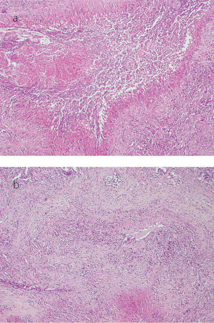
Figure 2. a, b.

Histopathologic specimens show fibrinoid necrosis with histiocytes and giant cells (a). There are lymphohistiocytic infiltrates which disturb the integrity of medium-sized vessels (b) [hematoxylin and eosin stain (H&E), original magnification ×100, ×200, respectively]
