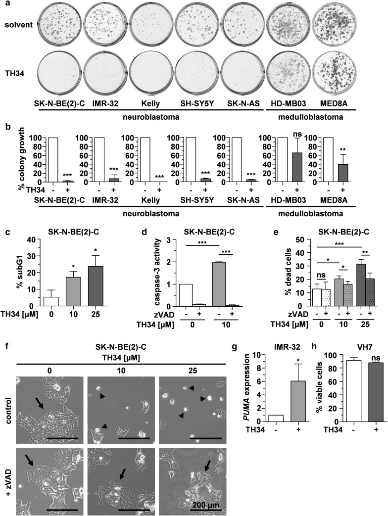
Fig. 2.
TH34 induces caspase-dependent programmed cell death in neuroblastoma cells. a, b Colony growth after treatment with TH34 (25 µM) or solvent. Representative images and quantification of colony growth in at least three independent experiments performed in triplicates are shown for each cell line. c Fraction of cells in subG1 cell cycle phase after treatment with indicated concentrations of TH34 for 72 h, identified via flow-cytometric quantification of DNA content using propidium iodide. d Caspase-3 activity after treatment of SK-N-BE(2)-C cells with indicated concentrations of TH34 for 48 h with or without Z-VAD-FMK (20 µM). e Proportion of dead SK-N-BE(2)-C cells after treatment with different concentrations of TH34 for 72 h with or without Z-VAD-FMK (20 µM), determined via automated trypan blue staining. f Representative images of SK-N-BE(2)-C neuroblastoma cells treated with solvent or TH34 (25 µM) with or without Z-VAD-FMK (20 µM) for 72 h. g Relative expression (determined using the 2−ΔΔCt method and normalized to solvent control) of PUMA in IMR-32 cells after 24 h of treatment with TH34 (10 µM). (h) VH7 non-malignant fibroblast viability after treatment with solvent or TH34 (25 µM) for 72 h. Bar graphs represent mean values of at least three independent experiments performed in triplicates and statistical analysis was performed using unpaired, two-tailed t test (***p < 0.001; **0.001 ≤ p < 0.01; *0.01 ≤ p < 0.05, ns not significant). Error bars represent SD

