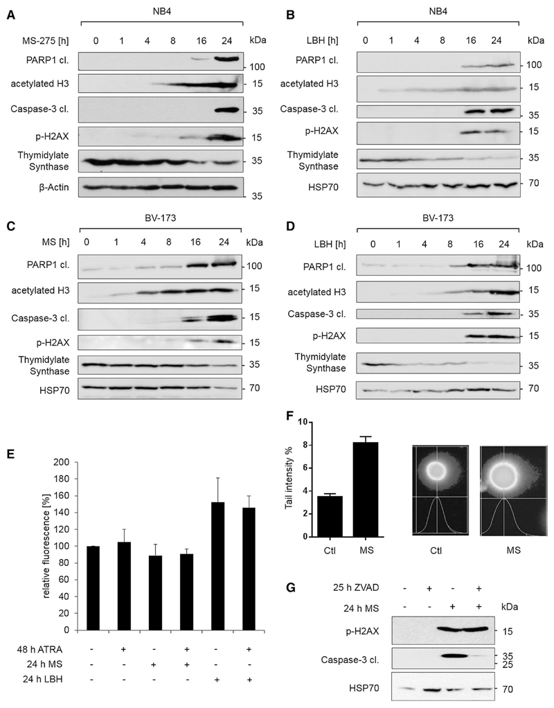
Fig. 4. HDACi induce DNA damage.
a–d NB4 and BV-173 cells were stimulated as indicated with 5 µM MS-275 (MS) or 100 nM LBH589 (LBH). Lysates were analyzed by immunoblot as indicated; n = 3. e NB4 cells were stimulated for the indicated times with 1 µM ATRA, 5 µM MS-275, or 100 nM LBH589 (LBH), and cytosolic ROS was measured. Unstained control was subtracted from each measurement, and data were normalized to control; n = 2. f NB4 cells were incubated for 12 h with 2 µM MS-275 and analyzed for DNA damage with comet assay; n = 2. g NB4 cells were treated as indicated and analyzed by immunoblot; 50 µM ZVAD-FMK (ZVAD) and 5 µM MS-275 were used. Different membranes with the same samples were used, n = 3

