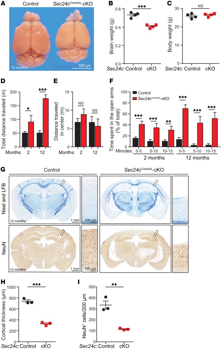
Figure 5. Postnatal deletion of Sec24c in the forebrain leads to hyperactivity and altered anatomy in the forebrain.
(A) Representative image of fixed brains dissected from a 12-month-old Sec24cCamk2a-cKO mouse and littermate control. Scale bar: 500 μm. (B and C) Brain (B) and body (C) weights (mean ± SEM) of 12-month-old Sec24cCamk2a-cKO and control mice (n = 4 mice/genotype). (D–F) Behavioral tests were performed using 2-month-old Sec24cCamk2a-cKO mice (n = 8) and age-matched controls (n = 17) and 12-month-old Sec24cCamk2a-cKO mice (n = 4) and age-matched controls (n = 5). Total distance traveled (mean ± SEM) (D) and distance traveled in the center (mean ± SEM) (E) by mice subjected to the open field test. (F) Mean time (± SEM) spent in the open arms during the indicated intervals by mice subjected to the elevated plus maze test. (G–I) Brain sections from 12-month-old Sec24cCamk2a-cKO and littermate control mice (n = 3 mice/genotype) were stained with Nissl and Luxol fast blue or immunostained with anti-NeuN antibody. Representative images (G), mean cortical thickness (± SEM) (H), and mean neuronal numbers (± SEM) (I) are shown. (G) Scale bars: 1 mm and 100 μm (insets). LFB, Luxol fast blue. *P < 0.05, **P < 0.01, and ***P < 0.001, by Student’s t test.

