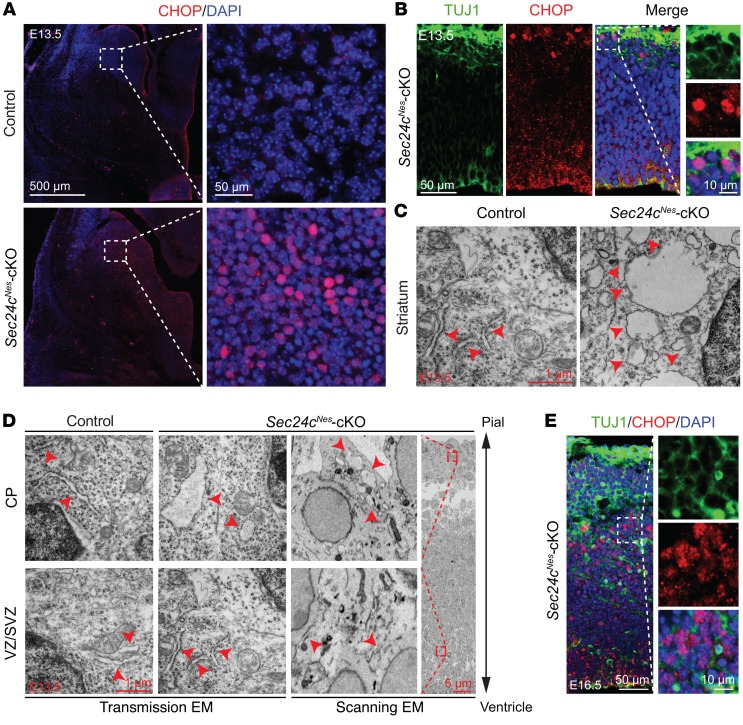
Figure 6. Sec24c deficiency causes ER stress.
(A) Representative images of immunostaining against CHOP in the striatum of control (n = 3) and Sec24cNes-cKO (n = 3) mice at E13.5. Scale bars: 500 μm and 50 μm (insets). (B) Representative images of CHOP staining in the cortex of Sec24cNes-cKO (n = 3) mice at E13.5. Scale bars: 50 μm and 10 μm (insets). (C) Representative ultrastructural images of E13.5 striatal neurons showing a swollen ER (denoted by red arrowheads) in Sec24c-deficient brain (n = 1). Scale bar: 1 μm. (D) Scanning and transmission EM analyses of the ultrastructure in cells located in the CP and the VZ/SVZ in Sec24cNes-cKO mice (n = 1). The ER structure is highlighted by arrowheads. Scale bars: 1 μm and 5 μm (inset). (E) Representative images of CHOP staining in the cortex of Sec24cNes-cKO mice at E16.5 (n = 3). Scale bars: 50 μm and 10 μm (insets).

