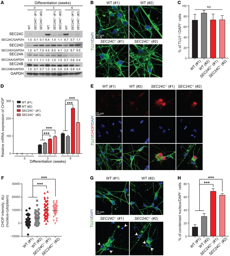
Figure 7. Deletion of SEC24C leads to elevated cellular stress and the demise of mature neurons derived from hiPSCs.
(A) Immunoblot analysis of cell extracts from WT and SEC24C-KO hiPSCs (clones 1 and 2) at 0, 3, and 6 weeks of neuronal differentiation. (B) Representative images of TUJ1 immunostaining of WT and SEC24C-KO hiPSCs at 6 weeks of differentiation. Scale bar: 10 μm. (C) Percentage of TUJ1+ cells per total cells (DAPI+) (mean ± SEM) shows normal differentiation of the SEC24C-KO hiPSCs compared with WT clones 1 and 2. (D) mRNA levels of CHOP in WT and SEC24C-KO hiPSCs at 0, 3, and 6 weeks of differentiation were determined by real-time RT-PCR. Data are presented as the mean ± SEM. (E) Representative images of immunostaining show pronounced nuclear localization of CHOP in SEC24C-KO hiPSCs at 6 weeks of differentiation. Scale bar: 10 μm. (F) Quantification of nucleus minus cytoplasm CHOP intensity shows significant enrichment of nuclear CHOP in SEC24C-KO hiPSCs. (G) Representative nuclear staining with DAPI and (H) quantification indicate a significant increase in the percentage of cells with condensed nuclei (mean ± SEM) in SEC24C-KO hiPSCs. Scale bar: 10 μm. Data were collected from 3 independent experiments. ***P < 0.001, by 2-way ANOVA.

