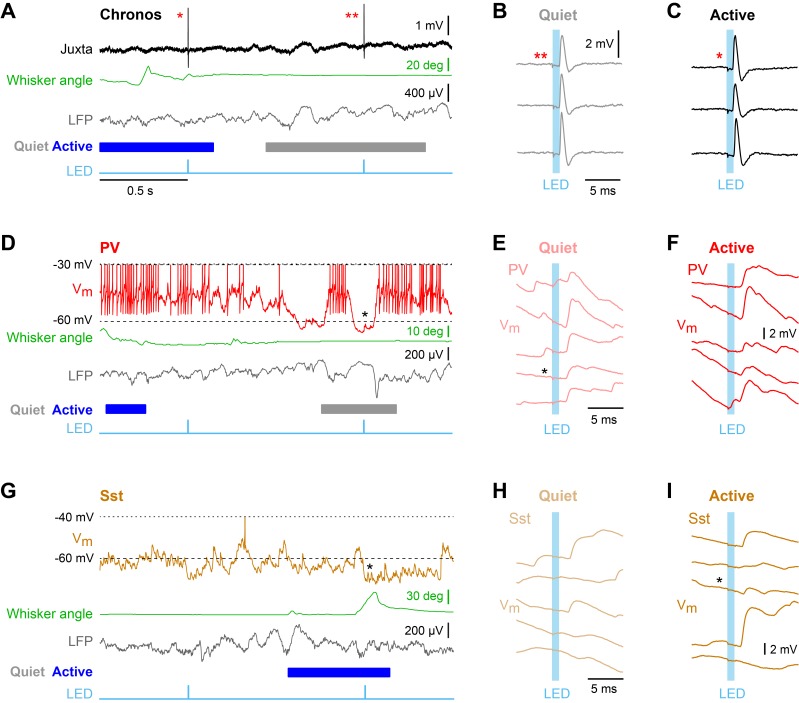
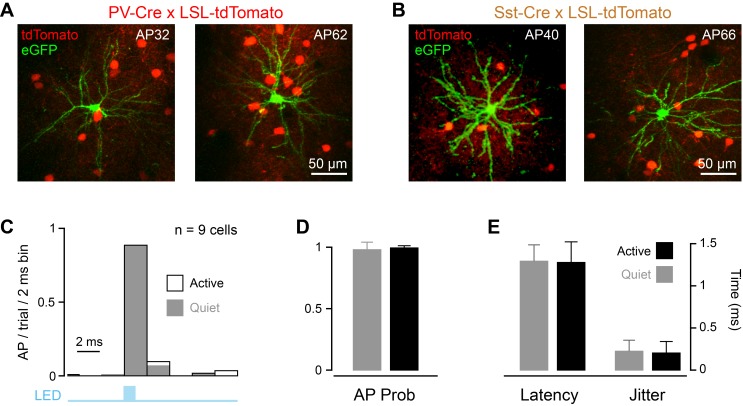
Figure 3. Unitary excitatory postsynaptic potentials in PV and Sst neurons in layer 2/3 of the awake mouse barrel cortex measured together with whisker position and local field potential.
(A) Example juxtacellular recording of a presynaptic Chronos-expressing neuron. From top to bottom: Extracellular signal (Juxta), whisker angle, LFP, and light stimulus (LED). Blue/grey color boxes represent Active and Quiet states. (B and C) Time-locked individual APs evoked by a 1 ms LED stimulus during Quiet and Active states. (D) Example whole-cell recording from a PV neuron together with whisker angle, LFP and LED stimulus. (E and F) Individual uEPSP responses to 1 ms optogenetic stimuli during Quiet and Active states. (G–I) Same as panels D-F, but for a Sst neuron.


