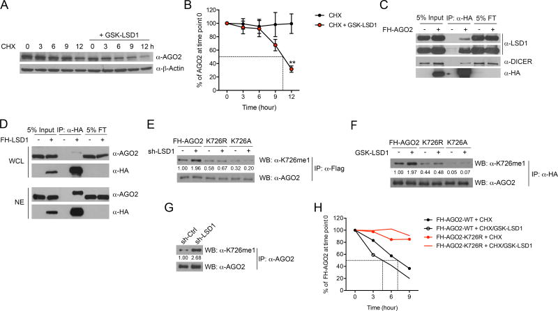
Figure 3. LSD1 regulates AGO2 methylation status and stability.
(A and B) Representative immunoblot of AGO2 (A) and quantification of AGO2 signal from five experiments (B, mean±SEM) in MCF-7 cells treated with 50 µg/ml cycloheximide (CHX) in the presence or absence of 2 µM GSK-LSD1 for the indicated times.
(C and D) The physical interaction between LSD1 and AGO2 was examined by co-IP assay using whole cell lysate (WCL) of MCF-7 cells stably expressing FH-AGO2 (C), or reciprocally using WCL and nuclear extract (NE) of MCF-7 cells stably expressing FH-LSD1 (D). FT-flow through.
(E and F) Ectopically expressed WT FH-AGO2 and mutants in MCF-7 cells treated by LSD1 KD (E) or GSK-LSD1 (F) were immunoprecipitated, and then immunoblotted with mono-methyl AGO2 specific antibody and an AGO2 antibody.
(G) The immunoblot of K726me1 on endogenous AGO2 in control or LSD1 KD MCF-7 cells.
(H) The protein stability of transiently expressed WT FH-AGO2 and FH-AGO2-K726R in 293T cells was measured using CHX chase assay in the presence or absence of 2 µM GSK-LSD1. The averaged AGO2 quantification from two experiments is shown.
**p < 0.01, as determined by unpaired t-test.
Also see Figure S3.

