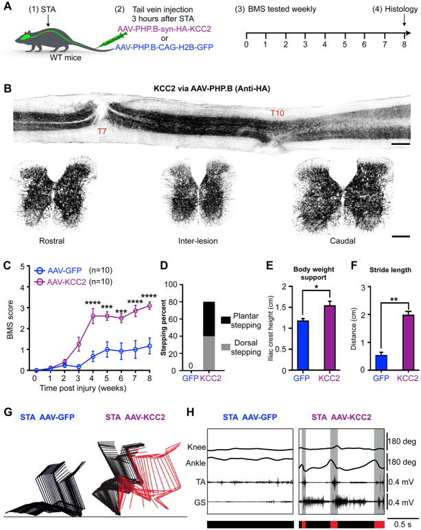
Figure 2. Widespread KCC2 expression mimics the effects of CLP290 to promote functional recovery.
(A) Experimental scheme.
(B) Representative image stacks of longitudinal (upper) and transverse (lower) spinal cord sections, taken from the mice at 8 weeks after staggered injury, stained with anti-HA (to detect the HA-KCC2 protein). Scale bar: 500 μm (upper) and 100 μm (lower).
(C) BMS performance in experimental (AAV-PHP.B-HA-KCC2) and control (AAV-PHP.B-H2B-GFP) groups. Two-way repeated-measures ANOVA followed by post hoc Bonferroni correction. *p < 0.05.
(D) Percentage of mice that reached stepping at 8 weeks after injury.
(E and F) Quantification of bodyweight support (E) and stride length (F) at 8 weeks (n = 10 per group). Student’s t-test (two-tailed, unpaired) was applied. *p < 0.05; **p < 0.01. Error bars: SEM.
(G) Color-coded stick view decomposition of mouse right hindlimb movement during dragging (AAV-PHP.B-H2B-GFP group) and stepping (AAV-PHP.B-HA-KCC2 group).
(H) Representative right hindlimb knee and ankle angle oscillation trace and simultaneous EMG recording of mice at 8 weeks after injury. Red bars, swing and stepping; black bars, stance and dragging.
See also Figure S4 and Supplementary Movie.

