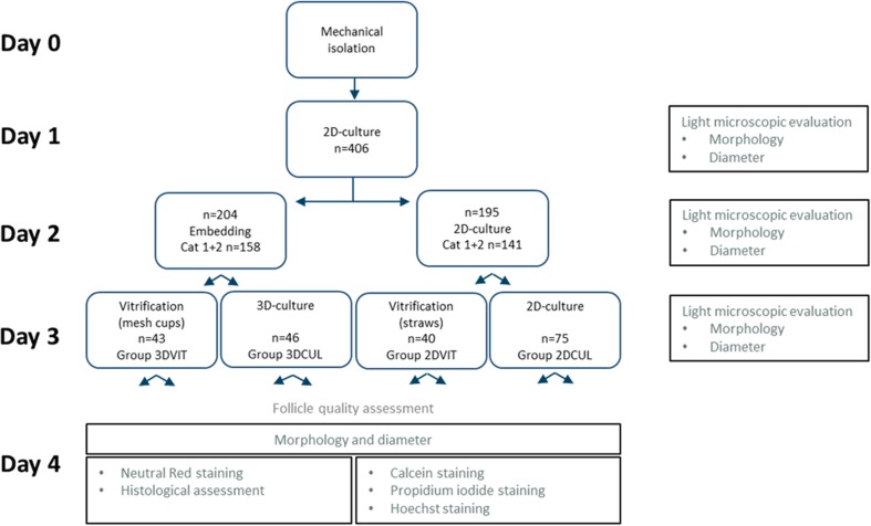
Fig. 2.
Experimental design (divided over 6 replicates). On each evaluation day (1, 2, 3, 4), morphology was assessed and follicle diameter measured. On day 4, half of the follicles from each group was stained with neutral red (NR) for viability assessment and fixated for histological assessment. The other half was used for calcein, propidium iodide and Hoechst staining. Grey text describes follicle quality assessment methods

