Figure 2.
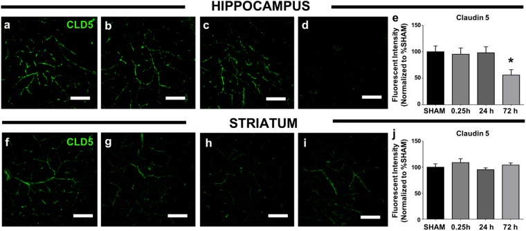
Claudin-5 expression is disturbed in brain regions vulnerable to blast-induced BBB disruption. (a) Shows representative images of CLD5 immunofluorescence (green) in sham hippocampal CA1, (b) at 0.25 h, (c) 24 h, and (d) 72 h after 2X blast. (e) Fluorimetric quantitation conducted by a condition-blind rater revealed a significant decrease in CLD5 fluorescence measured at 72 h after 2X blast. (f) Shows representative images of sham CLD5 immunofluorescence in dorsal striatum, (g) at 0.25 h, (h) 24 h, and (i) 72 h after 2X blast. (j) Fluorimetric quantitation revealed no significant differences in CLD5 fluorescence after 2X blast. One-way ANOVA post hoc Dunnett’s for CLD5. Values represent mean ± SEM; n = 4. (*p ≤ 0.05). Scale bars = 50 µm.

