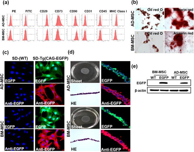
Figure 1.
Fabrication of rat mesenchymal stem cell sheets. (a) Histogram of cell surface markers showing the AD-MSC (upper) and BM-MSC (lower). (b) Differentiation capacity of AD-MSC and BM-MSC for differentiating into adipocytes and osteoblasts with alizarin red. (upper panel; undifferentiated condition, lower panel; differentiated condition). Extracellular calcium deposition in the osteoblasts is shown in red, the accumulation of lipid vacuoles in the adipocytes are shown in red. (c) EGFP signal of SD-Tg AD-MSC and BM-MSC 1 day after seeding on a culture dish. AD-MSC and BM-MSC were used at passages 3. (d) Fabricated AD-MSC and BM-MSC sheets. AD-MSC and BM-MSC sheets by hematoxylin-eosin staining. EGFP-expressing monolayered AD-MSC and BM-MSC sheets. AD-MSC and BM-MSC were used at passages 3 or 4. (e) EGFP protein expression levels in Tg-rat isolated AD-MSC and BM-MSC. All the experiments were performed three times. Green: AD-MSC or BM-MSC from EGFP transgenic rats, Red: EGFP antibody labeled by Alexa Flour 594 Blue: nucleus.

