Fig. 1.
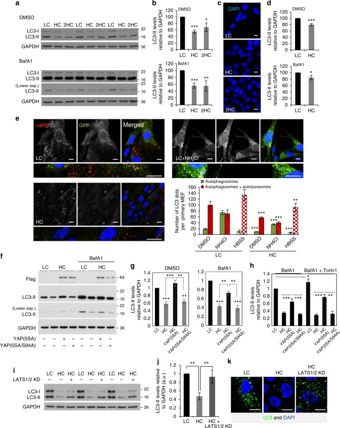
Autophagosome formation is reduced at high cell density via YAP/TAZ inhibition. a LC3-II levels assessed by immunoblotting in MCF10A cells plated at different confluencies: LC (low confluency) and HC (high confluency). 2HC – twice as many cells plated as in HC. The cells were treated with vehicle (DMSO) or bafilomycin A1 (BafA1) at 400 nM for 5 h. GAPDH was used as loading control. b Densitometry of LC3-II/GAPDH blots obtained from MCF10A cells plated at different confluencies as in a. The graphs show the mean ± s.d. (n = 3; ***P < 0.001, **P < 0.01, *P < 0.05; two-tailed one sample t-test). c Representative images of DAPI staining at different confluencies of MCF10A cells, used in a, b. Scale bar is 10 µm. d Densitometry of LC3-II/ Gapdh levels in primary MEFs plated at different confluencies. The graphs show the mean ± s.d. (n = 3; ***P < 0.001, *P < 0.05; two-tailed one sample t-test). e Representative confocal images (see left panel) and total number of GFP dots (autophagosomes) and mRFP dots (autophagosomes and autolysosomes)—right panel, in primary MEFs from transgenic mice expressing mRFP-GFP-LC3. MEFs plated at LC and HC were fixed and subjected to confocal visualization. The numbers of dots were counted automatically using a Cellomics microscope (n = 4; ***P < 0.001, **P < 0.01; two-tailed t-test). More than 600 cells were counted per experiment, per condition. Scale bar is 10 µm. f LC3-II levels assessed by immunoblotting in HeLa cells plated at different confluencies and overexpressing control (Ctrl), YAP(5SA) or YAP(5SA/S94A). These cells were exposed to either control (DMSO) or BafA1 (400 nM), for the last 5 h. GAPDH was used as loading control. g LC3-II/GAPDH densitometry of HeLa cells treated as in f. The graphs show the mean ± s.e.m. (n = 6; ***P < 0.001, **P < 0.01; two-tailed one sample t-test). h LC3-II/GAPDH densitometry of HeLa cells overexpressing either YAP(5SA) or YAP(5SA/S94A) in the presence of BafA1 and/or Torin1 (used as mTORC1 inhibitor). GAPDH was used as loading control. Bars represent the mean ± s.d. (n = 3; ***P < 0.001, **P < 0.01, *P < 0.1; two-tailed t-test). i LC3-II levels assessed by immunoblotting in MCF10A cells plated at different confluencies and exposed to either control (Ctrl) or LATS1/2 siRNAs. GAPDH was used as loading control. j LC3-II/GAPDH densitometry of MCF10A treated as in h. The graphs show the mean ± s.d. (n = 4; **P < 0.01; two-tailed one sample t-test). k Representative images of endogenous imunostaining of LC3 in MCF10A cells treated as above. Quantification of number of LC3 dots per cell and enlarged images are presented in Supplementary Fig. 4b. n = number of independent biological replicates unless otherwise stated

