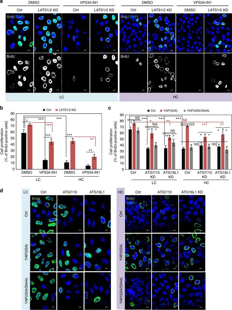
Fig. 8.
The role of autophagy on YAP/TAZ regulation of cell proliferation. a Representative BrdU images of MCF10A cells exposed to LATS1/2 siRNAs, plated at low and high densities and treated with VPS34-IN1 (1 µM) for the last 24 h. Scale bars are 10 µm. b Quantification of MCF10A cells positive for BrdU (cell proliferation). MCF10A cells were treated as in a. Bars represent the mean ± s.d. (n = 3; ***P < 0.001, **P < 0.01; two-tailed t-test). c Quantification of HeLa cells positive for BrdU (cell proliferation). HeLa cells were initially transfected with control, ATG7/ATG10 or ATG16L1 siRNAs, then transfected with control (empty flag), YAP(5SA) or YAP(5SA/S94A) constructs and plated at low and high confluencies. Bars represent the mean ± s.d. (n = 3; ***P < 0.001, **P < 0.01, *P < 0.05, NS not significant; two-tailed t-test). d Representative BrdU images of HeLa cells overexpressing control (empty flag), YAP(5SA) or YAP(5SA/S94A) for the quantification in c. The cells were initially exposed to control, ATG7/ATG10 or ATG16L1 siRNAs. Scale bars are 10 µm. n = number of independent biological replicates unless otherwise stated

