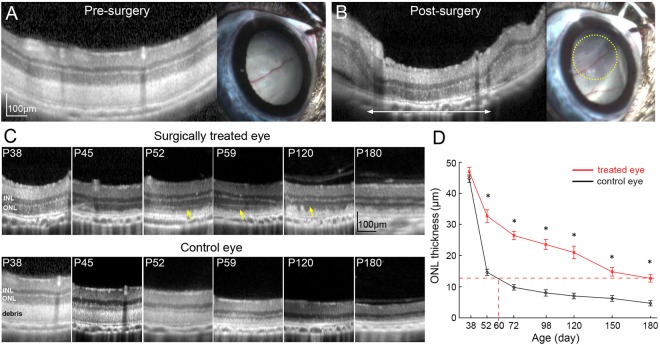
Figure 1.
Removal of subretinal debris in RCS rats preserves photoreceptors. (A) OCT and fundus image before surgery. (B) After the surgery, debris-free area in OCT image is outlined with a white arrow. Retina became more transparent on fundus photography (within the yellow dotted line). (C) Serial OCT scans demonstrate better preservation of photoreceptors in treated eyes, as compared to control eyes. Photoreceptor outer segments reappear a few days after debridement (yellow arrows). (D) ONL is significantly thicker in the treated area than in the corresponding controls. In treated eyes, the ONL thickness at P180 is equivalent to that of P60 in controls (n = 10 control eyes and 10 treated eyes in 10 animals, error bars - s.e.m., *p < 0.01, two-sided paired t-test, p-values respectively 0.36, 2.5E-05, 8.2E-07, 2.2E-05, 0.00023, 0.0014, 5.8E-05).

