Figure 4.
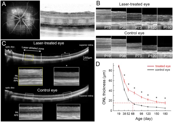
Preservation of retinal structure after laser photocoagulation. (A) Laser was applied to the central retina around the optic nerve head at P19. Pattern of photocoagulation can be seen in fluorescein angiography and in OCT. (B) Laser treatment delays the loss of photoreceptor nuclei from P19 through P180, compared to control eyes. (C) ONL was much thicker in laser-treated retina (yellow box) compared to untreated periphery (white box with asterisk). Nearly no ONL could be seen in the corresponding zones of the control eye (lower two white boxes) (P98). (D) Significantly higher ONL thickness in the laser-treated retina, compared to the corresponding areas of the control eye. In treated eyes, the ONL thickness at P180 is equivalent to that of P68 in control eyes. (n = 8 control eyes and 8 treated eyes in 8 animals, error bars - s.e.m., *p < 0.05, two-sided paired t-test, p-values respectively 0.29, 4.7E-04, 7.1E-04, 1.2E-05, 8.3E-06, 5.6E-04, 8.1E-04, 0.011).

