Fig. 1.
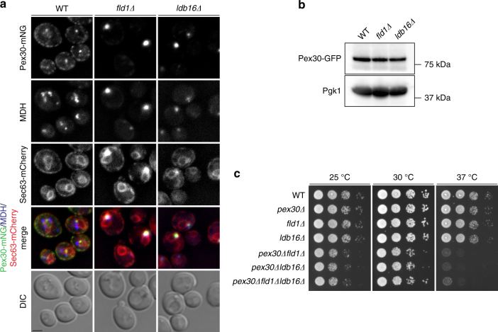
Pex30 is localized to LD proximal regions in seipin mutants. a Localization of endogenous Pex30 tagged with monomeric Neon Green (Pex30-mNG) in WT, fld1∆, and ldb16∆ cells. Endogenous Sec63 was tagged with mCherry and used as an ER marker. LDs were stained with the neutral lipid dye MDH. Bar 2 µm. b Levels of endogenous GFP-tagged Pex30 in WT, fld1∆, and ldb16∆ cells. Whole-cell extracts were separated by SDS-PAGE and analyzed by western blotting. Pex30-GFP and Pgk1, used as loading control, were detected with anti-GFP and -Pgk1 antibodies, respectively. c Tenfold serial dilutions of cells with the indicated genotype were spotted on YPD media and incubated at 25 °C, 30 °C, or 37 °C for 2 days

