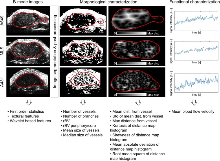
Figure 1.
Image biomarker extraction from A549, MLS and A431 tumours. B-mode images were used to calculate first order statistics, textural features and wavelet-based features of the tumours. Morphological features characterizing the tumour vasculature were extracted from an automated blood vessel segmentation algorithm and its corresponding distance map. The scale bars correspond to 1 mm. Functional characteristics of the tumour vasculature were obtained by using the time intensity curves of the acquired contrast-enhanced ultrasound data.

