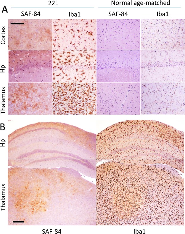
Figure 1.
Immunohistochemistry of 22L-infected mouse brains. (A) Large magnification images of PrPSc deposition stained with SAF-84 antibody and activated microglia stained for Iba1 in cortex, hippocampus (Hp) and thalamus. SAF-84 and Iba1 staining of uninfected, age-matched controls is shown as references. Scale bar = 100 μm. (B) Low magnification images of PrPSc deposition stained with SAF-84 antibody and activated microglia stained for Iba1 in hippocampus (Hp) and thalamus. Scale bar = 300 μm.

