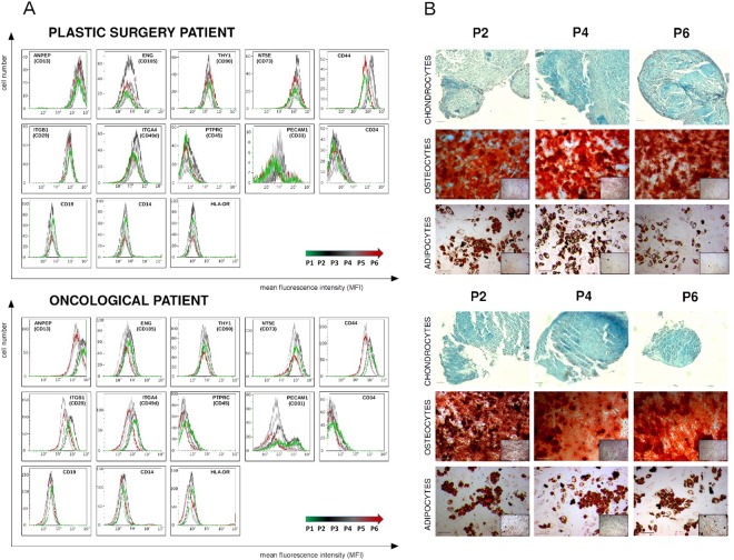
Figure 2.
Phenotype identification (A) and assessment of differentiation potential (B) of ASCs originating from plastic and oncological surgery patients. (A) Phenotyping of ASCs standard and supplementary markers using flow cytometry (FCM). FCM plots are shown for two representative patients: plastic and oncological surgery. The X axis represents mean fluorescence intensity (MFI), Y axis - cell number. (B) Representative example of histochemical analysis of ASCs differentiation into chondrocytes, osteocytes and adipocytes from plastic surgery and oncological patients. Differentiation assays were performed after 2nd (P2), 4th (P4), and 6th (P6) passage, oil red O, alizarin red S and 1% alcian blue staining were used to confirm adipocytes, osteocytes and chondrocytes differentiation respectively.

