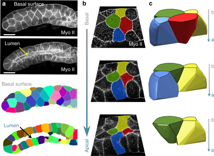
Fig. 2.
3D tissue packing of curved epithelia. a Example of Drosophila salivary gland and its processed images. Scale bar = 100 μm. b Confocal images showing the apico-basal cell intercalation of epithelial cells marked with green, yellow, red, and blue pseudo-colours. The green cell participates in two apico-basal transitions. c 3D reconstruction of the same cells labelled in b using the same colour code. The image confirms the presence of concave surfaces predicted by the mathematical model

