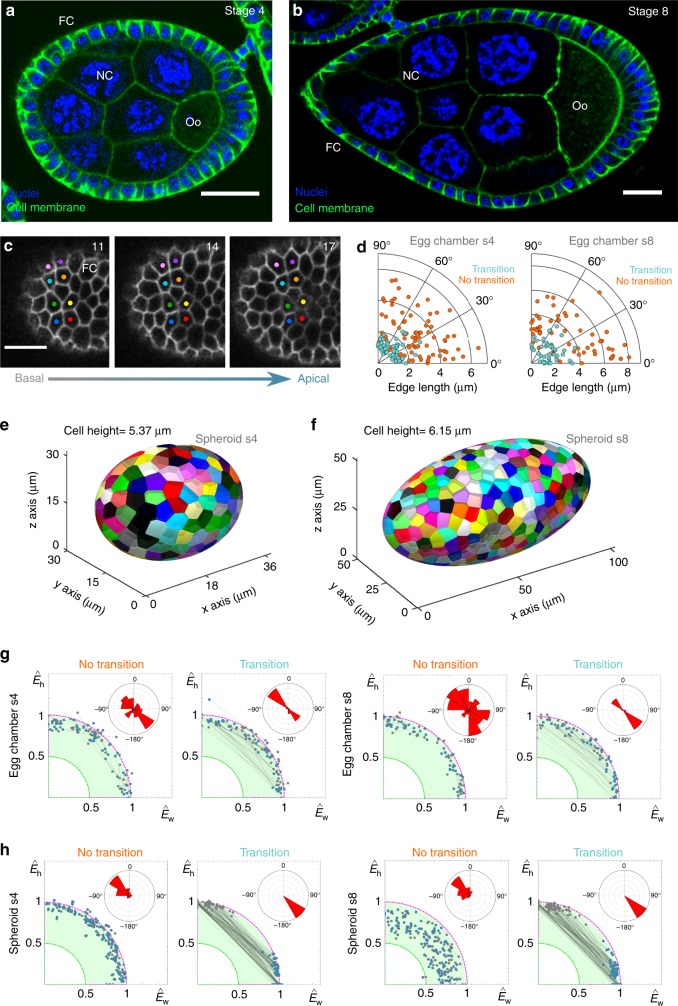
Fig. 5.
Graphic summary of 3D packing in epithelia. a Section of a stage 4 egg chamber with nuclei (blue) and cell membranes (green) labelled. The green staining decorates the contours of the follicular cells (FC), the nurse cells (NC) and the oocyte (Oo). Scale bar = 20 μm. b Section of a stage 8 egg chamber with nuclei (blue) and cell membranes (green) labelled. The green staining decorates the contours of the follicular cells (FC), the nurse cells (NC) and the oocyte (Oo). Scale bar = 20 μm. c Close up of the surface of a stage 4 egg chamber (a), showing two four-cell motifs that are involved in an apico-basal transition. The numbers indicate the confocal plane, increasing from basal to apical regions of the follicular cells. In this case, the sections were taken every 0.4 μm. Scale bar = 10 μm. d Polar scatters showing the length and the angle of the contacting edge in basal four-cell motifs from the stage 4 and stage 8 egg chambers. Light blue points stand for motifs that exchange neighbours; orange points stand for motifs that do not intercalate. e Voronoi spheroid model with the same dimensions that the average egg chamber 4. f Voronoi spheroid model with the same dimensions that the average egg chamber 8. g–h Linear decomposition of the experimental energies into the fundamental modes and . Colour codes as in Fig. 4

