Figure 3.

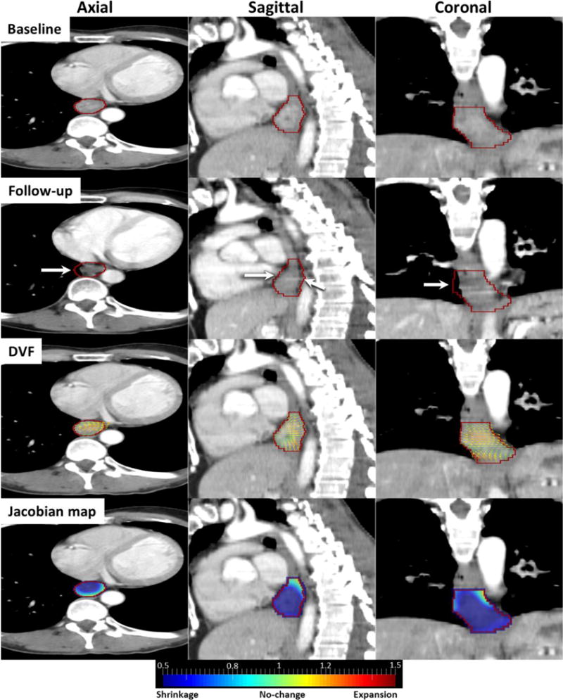
Responder case: Baseline, follow-up, DVF and Jacobian images in axial, sagittal and coronal views. Red contour is GTV and white arrows indicate shrinking esophageal wall.

Responder case: Baseline, follow-up, DVF and Jacobian images in axial, sagittal and coronal views. Red contour is GTV and white arrows indicate shrinking esophageal wall.