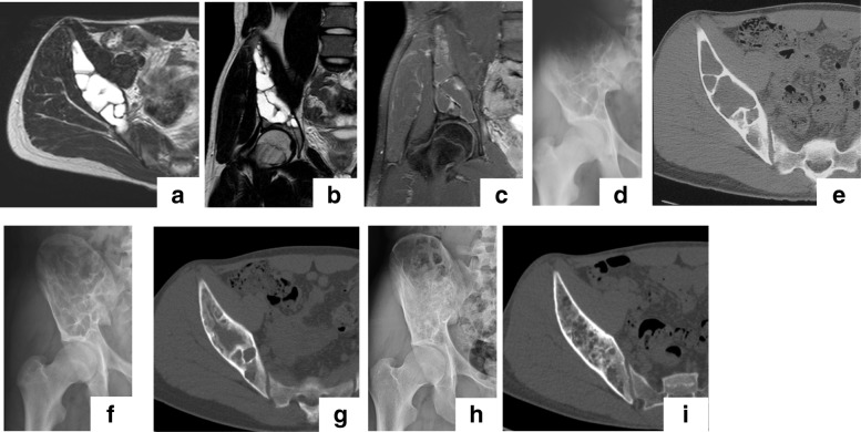
Fig. 4.
Typical case of a suspected ABC treated with ESC. Magnetic resonance (MR) images of a 16-year-old boy who presented with bruising of his hip sustained during contact sports participation: (a) axial T2; (b) coronal T2-weighted; and (c) coronal T1-weighted enhanced with gadolinium (Gd). The plain radiograph is shown in (d). The preoperative computed tomography (CT) image (e) shows the bone lesion with cortical thinning extending from the iliac wing to the periacetabular area. Plain radiographs after surgery (f). CT image at 6 months after ESC (g) shows remodeling of the cystic lesion by cancellous bone. At 6 years after ESC (h), the pelvic bone was fully remodeled despite a residual cyst and without any symptoms (class B) (i)

