Fig. 4.
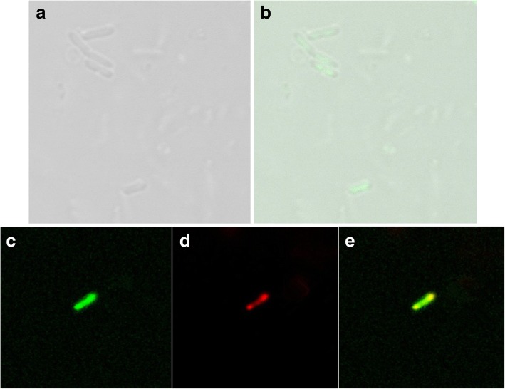
Localization of loaded DNA in L. Casei ghosts visualized by confocal laser scanning microscopy. a L. casei 393 ghosts filled with SYBR Green I labeled pCI-EGFP under white light. b L. casei ghosts filled with SYBR Green I labeled pCI-EGFP. Overlay of differential interference contrast and fluorescent image. The image indicated that the plasmid was filled within the bacterial ghosts. c Ghost membranes were stained with MitoTracker Green FM (green). d pCI-EGFP was detected by in situ hybridization with Cy3-labeled probes (red) specific for the EGFP. e All fluorescence photomicrographs (c, d) were taken of one middle z-scan section through the middle plane of the loaded bacterial ghosts and subsequently overlaid (e). Direct overlays of red and green fluorescent structures are represented by the yellow color

