
FIGURE 6.
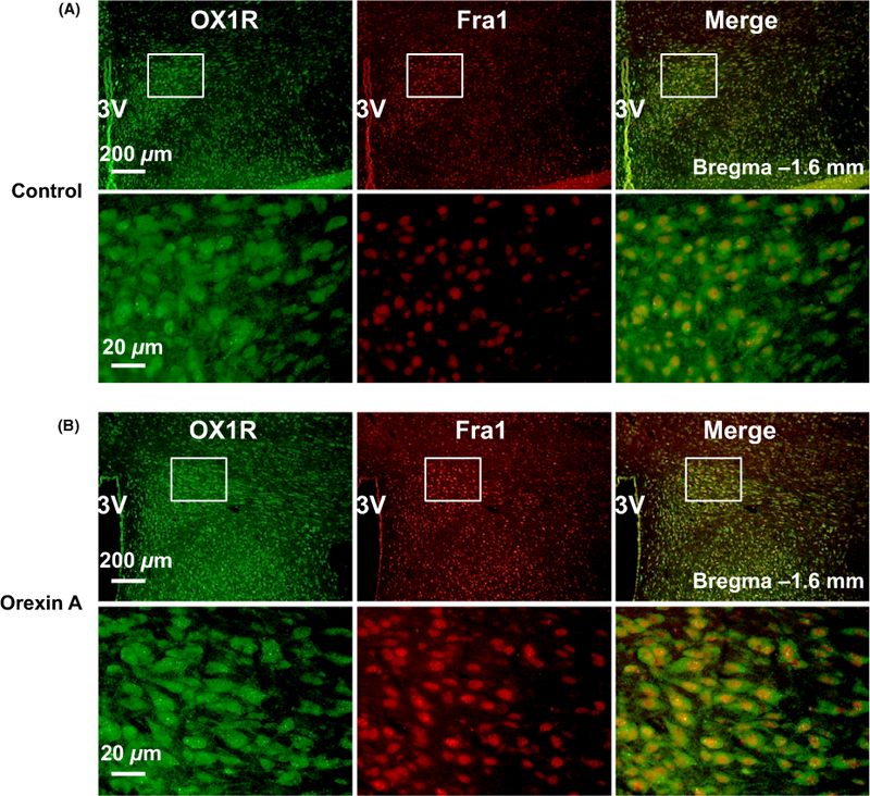
Colocalization of OX1R and Fra1 in the paraventricular nucleus (PVN). (A) a representative micrograph showing immunoreactivity of OX1R (green), Fra1 (red) and merged image in the PVN of vehicle control rat; (B) a representative micrograph showing immunoreactivity of OX1R (green), Fra1(red) and merged image in the PVN of central administration of orexin A SD rat. The brain coronal sections were taken from bregma −1.6 mm. 3V, the third ventricle