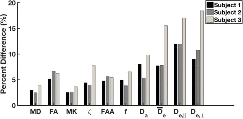
Figure 4.
Median absolute percent difference between Runs 1 and 2 for selected diffusion measures in white matter. These were calculated for all three subjects on a voxelwise basis by using all voxels considered as white matter (i.e., MK ≥ 1). In most cases, the percent difference is about 10% or less. However, the extra-axonal diffusivites ( , De,‖, De,⊥) differed by up to 20% for Subject 3. All quantities were obtained with either DKI (MD, FA, MK), FBI (ζ, FAA), or FBWM (f, Da, , De,‖, De,⊥).

