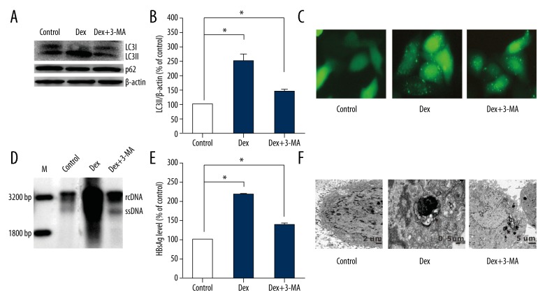
Figure 4.
Autophagy inhibitors (3-MA) decreases HBsAg secretion in HepG2.2.15 treated with dexamethasone (Dex). HepG2.2.15 cells were treated with 3-MA+ Dex and Dex respectively, after transfecting with GFP-LC3 plasmids for 24 h. (A) 36 h later, cells were collected and analyzed the conversion of LC3-I to LC3-II and p62 by western blot. (B) The LC3-II/β-actin ratios were quantified by densitometric analysis with Quantity One software (Bio-Rad). Results presented were representative of 3 independent experiments. (C, F) Immunofluorescence images for autophagic dots and TEM in pGFP-LC3-transfected hepG2.2.15 cells treated with 3-MA+ Dex and Dex. (D) HBV intermediates were extracted and subjected to southern blot analysis. (E) HBsAg secreted in the supernatants were analyzed by ELISA assay.

