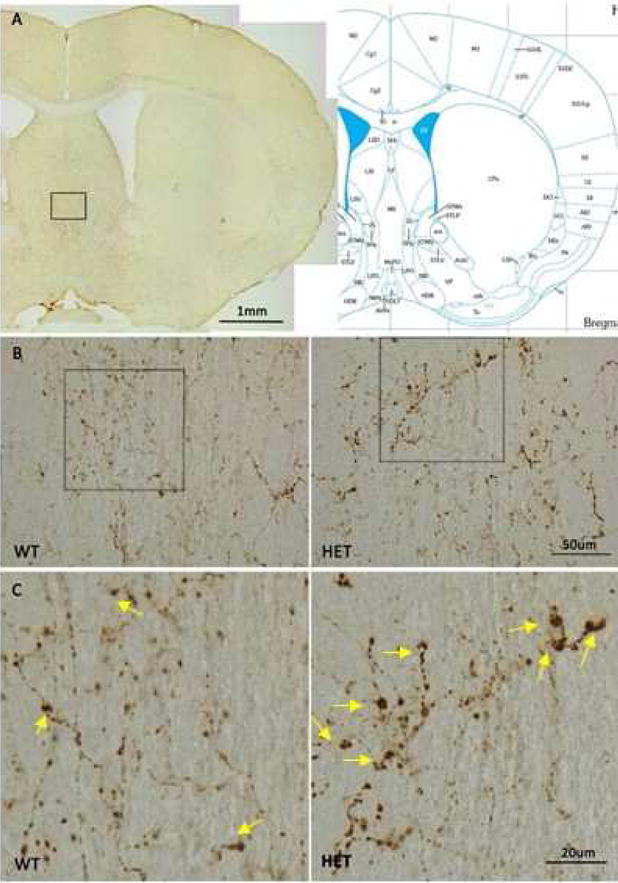
Figure 6.

A) Diagram showing medial septal brain region in study. B. Orexin positive aggregates at terminals in wild-type and heterozygous mice. Lower magnification (20×) shown in B, higher magnification (60×) shown in C. BiP heterozygous mice display many more enlarged terminals staining positive for orexin compared to wild-type mice. Arrows in the bottom panels) indicate the large varicosities.
