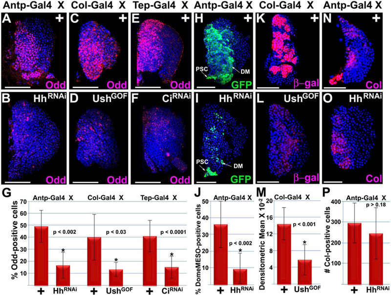
Figure 5. Hedgehog signaling maintains the Odd-positive and DomeMESO-positive but not Col-positive prohemocyte population.

(A–G) Disrupted Hh signaling significantly reduced the percentage of Odd-positive prohemocytes compared to controls. Hh signaling was disrupted by (A–D) reducing Hh expression in the PSC (Antp>HhRNAi or Col>UshGOF) or (E,F) loss of Ci function in the MZ (Tep>CiRNAi). (H–J) Knockdown of Hh expression in the PSC resulted in a significant reduction in the number of DomeMESO-GFP expressing prohemocytes. (K–M) Disrupting Hh signaling by mis-expressing Ush in the PSC (Col>UshGOF) significantly reduced DomeMESO-βgal expression levels. (N–P) Knockdown of Hh expression in the PSC did not significantly reduce the number of Col-positive prohemocytes. (G,J,M,P) Histograms show the results of the statistical analyses. Student’s t-test; error bars show standard deviation; P values are as shown; (G) control and HhRNAi (n=11), control and UshGOF (n=10), control and CiRNAi (n=10); (J) control and HhRNAi (n=11); (M) control and UshGOF (n=12); (P) control and HhRNAi (n=11). Mid-third instar lymph glands are counterstained with Dapi. Scale bars: 50 μm.
