
Fig. 1. Schematic of the mock screening protocol employed in this study.
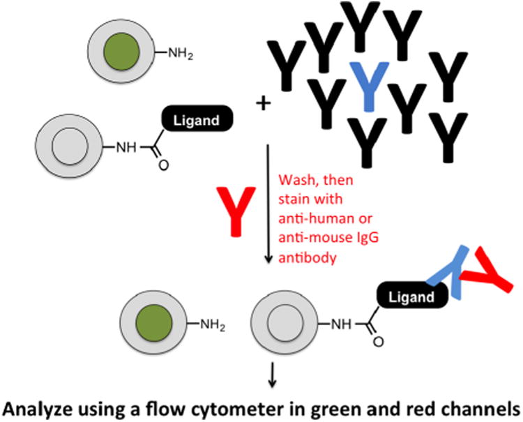
Cartoon of the mock screening strategy. “Blank beads” labeled internally with carboxyfluorescein were mixed with unlabeled beads displaying a known ligand to the antibody of interest at a ratio of 10:1. The monoclonal antibody targeted by the ligand (blue) was then doped into serum containing a multitude of IgG antibodies (black) at a known concentration. The beads and serum were mixed, washed, then stained with an Alexa Fluor 647 (A647)-conjugated secondary antibody. After a final wash the beads were analyzed by flow cytometry.