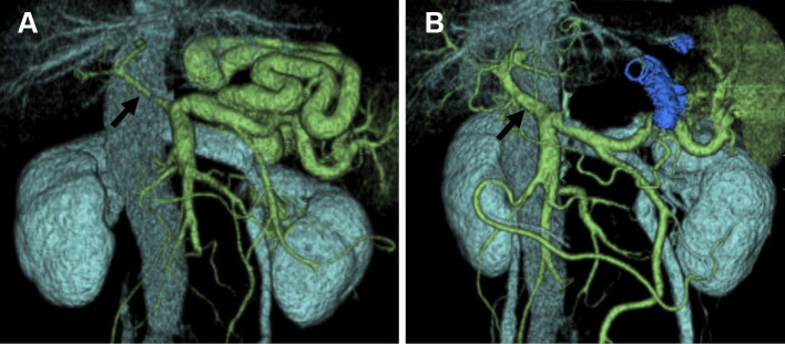
Figure 2.
(A) Three-dimensional computed tomography performed before balloon-occluded retrograde transvenous obliteration clearly showing a thick, long, and winding shunt supplied by the splenic vein and draining into the left renal vein. The main tract of the portal vein is markedly narrow (arrow). (B) Three-dimensional computed tomography one week after balloon-occluded retrograde transvenous obliteration showing complete occlusion of the spleno-renal shunt and an increase in the diameter of the main tract of the portal vein, suggesting an increased blood flow into the liver (arrow).

