
Figure 3.
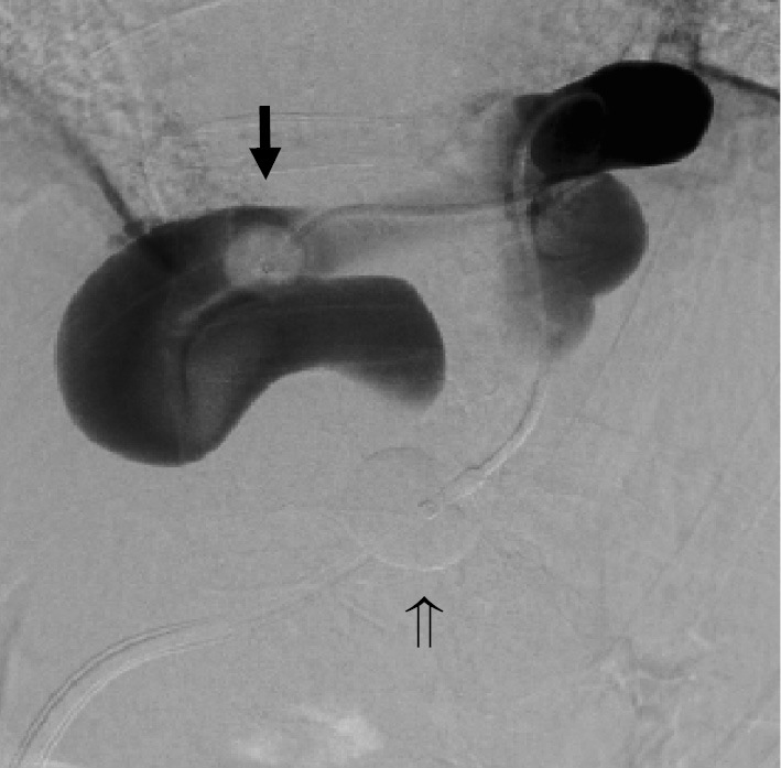
Balloon-occluded retrograde transvenous obliteration was attempted using a coaxial and double interruption system. Cytography was performed by inserting the catheter into the shunt. Complete balloon occlusion was not achieved with a 5-Fr balloon catheter (arrow ➡). The φ20 mm balloon was inflated when the guiding balloon catheter was fixed at the optimum position (arrow ⇒), following which stasis of the contrast medium was observed. We then performed balloon-occluded retrograde transvenous obliteration using a sclerosing agent.