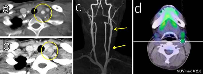
Figure 3.
Imaging of arteritis in the left carotid artery. A double ring pattern of wall thickening was observed on contrast computed tomography (a: arterial phase; b: venous phase). Magnetic resonance imaging revealed irregular narrowing of the left carotid artery (c). Positron emission tomography showed mild uptake in the left common carotid artery (d).

