Figure 1.

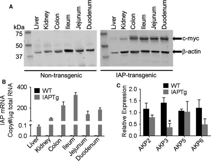
Villin promoter‐driven intestine‐specific expression of IAP. At 10 weeks of age nontransgneic (WT) and IAPTg littermates were euthanized and RNA as well as total protein extracts were prepared from the indicated tissues. (A) Total protein extracts from indicated tissues were analyzed by Western blot analyses using primary antibody to c‐myc tag and β‐actin. Representative blots are shown and immune‐reactive bands are indicated by arrows. (B) mRNA levels of chimeric IAP were quantified by QPCR as described under Methods and IAP mRNA copy number was calculated. (C) Total RNA from ileum was used to determine the mRNA levels of indicated endogenous IAP isoforms by QPCR. Data are expressed as Mean ± SD, n = 3. *P < 0.05.
