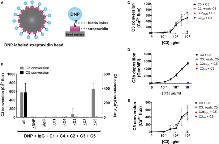
Figure 4.
Development of a classical pathway C3 and C5 convertase model. (A) In the CP convertase model, streptavidin beads are labeled with biotinylated DNP which serves as a model antigen. Addition of IgG1 recognizing DNP and complement proteins results in formation of C4b2a and C4b2a(C3b)n convertases on the bead surface, which convert C3 and C5. (B) Only in the presence of all CP components (antigen, IgG, and CP proteins) C3 and C5 are converted, as measured by calcium mobilization in U937-C3aR and U937-C5aR cells, respectively. (C) C3 conversion via CP convertases results in release of C3a in the supernatant (as shown by calcium mobilization) and (D) C3b deposition on the bead surface (as shown by flow cytometry). Identical amounts of the non-reactive C3 variants C3bH2O and C3MA do not bind to the bead surface. (E) C5 convertase activity increases with the level of deposited C3b molecules on the beads surface, but is not affected by C3bH2O or C3MA in solution. Uncoupling C3 and C5 conversion by introduction of an extra washing step does not alter C5 conversion, indicating that CP C5 conversion only depends on deposited C3b molecules around existing C3 convertases. (B–E) Data of three independent experiments, presented as mean ± SD.

