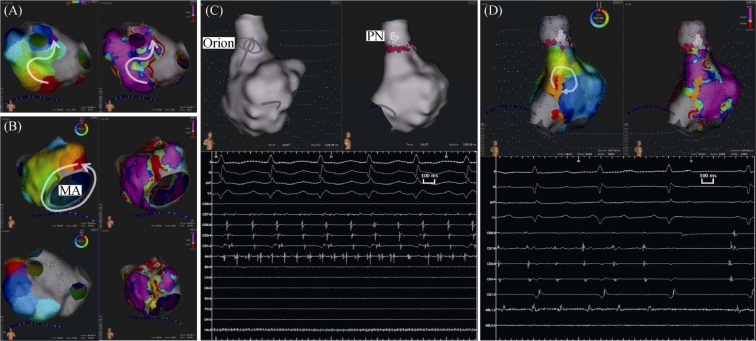
Figure 2. Conduction gap detection, AT induction and endocardial catheter ablation.
(A): conduction gap at rigid of left inferior PV during CS ostium pacing-rhythm; (B): LA activation mapping present counter clock-wise activation around MA during AT1 and ablation lesion was set at LA anterior wall; (C): SVC firing was detected and PN was marked, then ablation was performed for SVC isolation; (D): further RA activation mapping, micro re-entry activation pattern was present at RA lower crista terminalis during AT2. And AT2 was terminated soon during ablation. AT: atrial tachycardia; CS: coronary sinus; LA: left atrium; MA: mitral annuls; PN: phrenic nerve; PV: pulmonary vein; RA: right atrium; SVC: superior vena cava.

