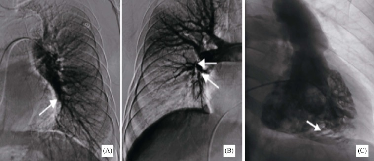
Figure 2. Pulmonary and right ventricular angiography.
(A) & (B): Pulmonary angiography: the left lower pulmonary artery exhibits thrombosis (left), and the middle and lower lobes of right pulmonary artery occur occlusion (right); (C): right ventricular angiography: the black part (arrow) is the retention of contrast agent, and the white part is the trabecular muscles.

