Fig. 3.
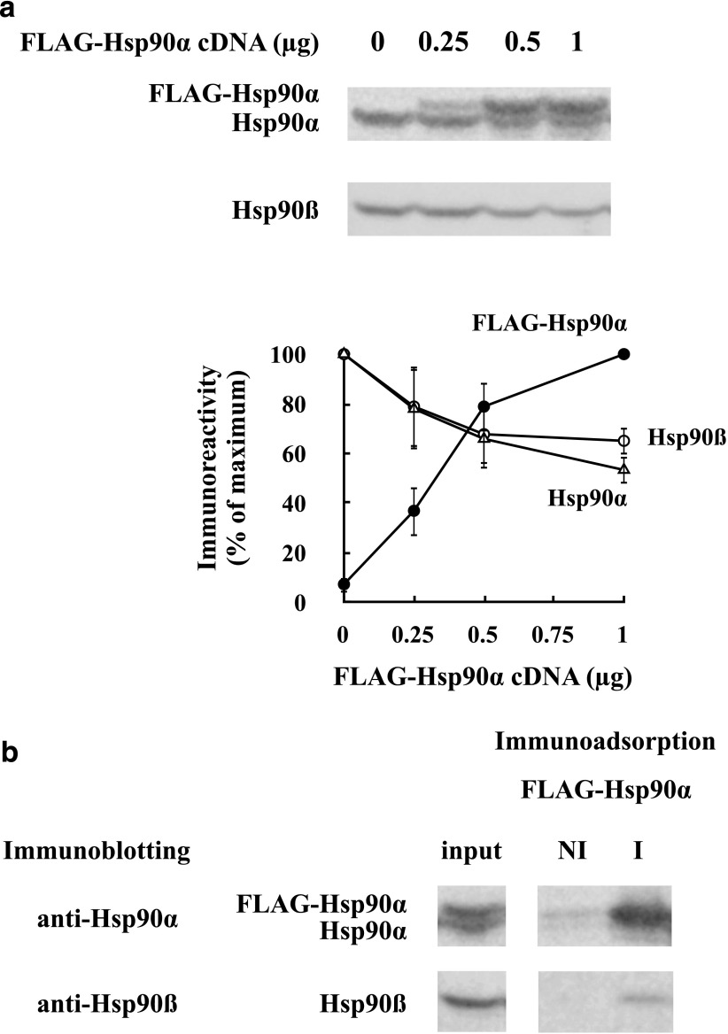
Expression of FLAG-Hsp90α in HEK cells suggests that the total amount of Hsp90 is under homeostatic regulation. (a) HEK cells were transfected with FLAG-Hsp90α cDNA, and 48 hours later, lysates were prepared and immunoblotted for Hsp90α and Hsp90β. The graph shows the mean ± S.D. of scans for three experiments expressed as the percentage of maximum immunoreactivity. (●) FLAG-Hsp90α, (△) endogenous HEK Hsp90α, (○) HEK Hsp90β. (b) FLAG-hHsp90α forms primarily homodimers with itself and not mixed dimers with endogenous HEK Hsp90α or Hsp90β. Lysate prepared as above was immunoadsorbed with anti-FLAG (I) or nonimmune (NI) antibody and immunoblotted with anti-Hsp90α or anti-Hsp90β.

