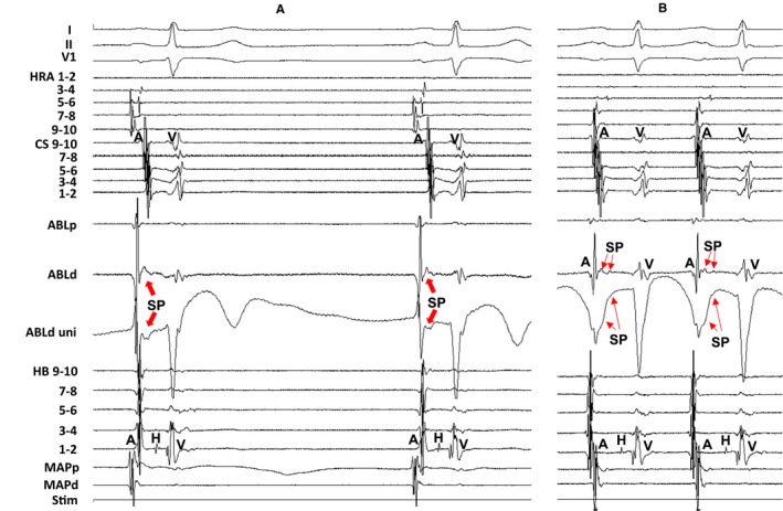
Figure 5.

Tracing during sinus rhythm (A) and during atrial tachycardia (B) at the successful ablation site in the same patient as in Figure 2. The amplitude of the slow potential (SP) was attenuated and the electrogram width of the SP was prolonged during atrial tachycardia (B) compared with the SP during sinus rhythm (A). The electrocardiographic leads I, II, and V1, and electrograms recorded at the high right atrium (HRA), coronary sinus (CS), ablation catheter (ABL), and His bundle (HB) position are shown. ABLd indicates distal site of ablation catheter; ABLp, proximal site of ablation catheter; MAPd, distal site of mapping catheter; MAPp, proximal site of mapping catheter; Stim, stimulation; uni, unipolar electrogram.
