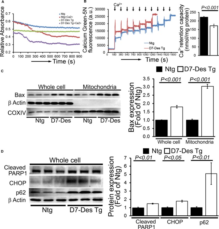
Figure 7.

Altered mitochondrial swelling and calcium retention capacity in D7‐Des Tg hearts. A, Representative images of calcium‐induced mitochondrial swelling isolated from the D7‐Des Tg and Ntg hearts at 6 months of age. Mitochondrial swelling was induced by the addition of 200 μmol/L CaCl2. The assay was performed in 3 independent experiments, and representative tracings are shown. B, Representative traces and quantification of mitochondrial Ca2+‐retention capacity of D7‐Des Tg and Ntg hearts at 6 months of age. Fluorescence reading of Ca2+ measured with Calcium Green‐5N indicator in solution with subsequent addition of Ca2+ pulses of 20 nmol/mg of mitochondrial protein. Cumulative Ca2+ additions are shown at each arrowhead (n=5–6 mice per group). C, Representative Western blot and densitometric quantification shows significantly increased Bax expression and mitochondrial localization in the D7‐Des Tg mice hearts. D, Representative Western blot and densitometric quantification showing increased expression of cleaved PARP1, CHOP, and p62 protein isolated from the D7‐Des Tg mice heart (n=6 mice per group). Bars represent mean±SEM. P values were determined by Tukey's post hoc test. D7‐Des Tg indicates mutant desmin transgenic mouse; Ntg, non‐transgenic.
