Figure 5.

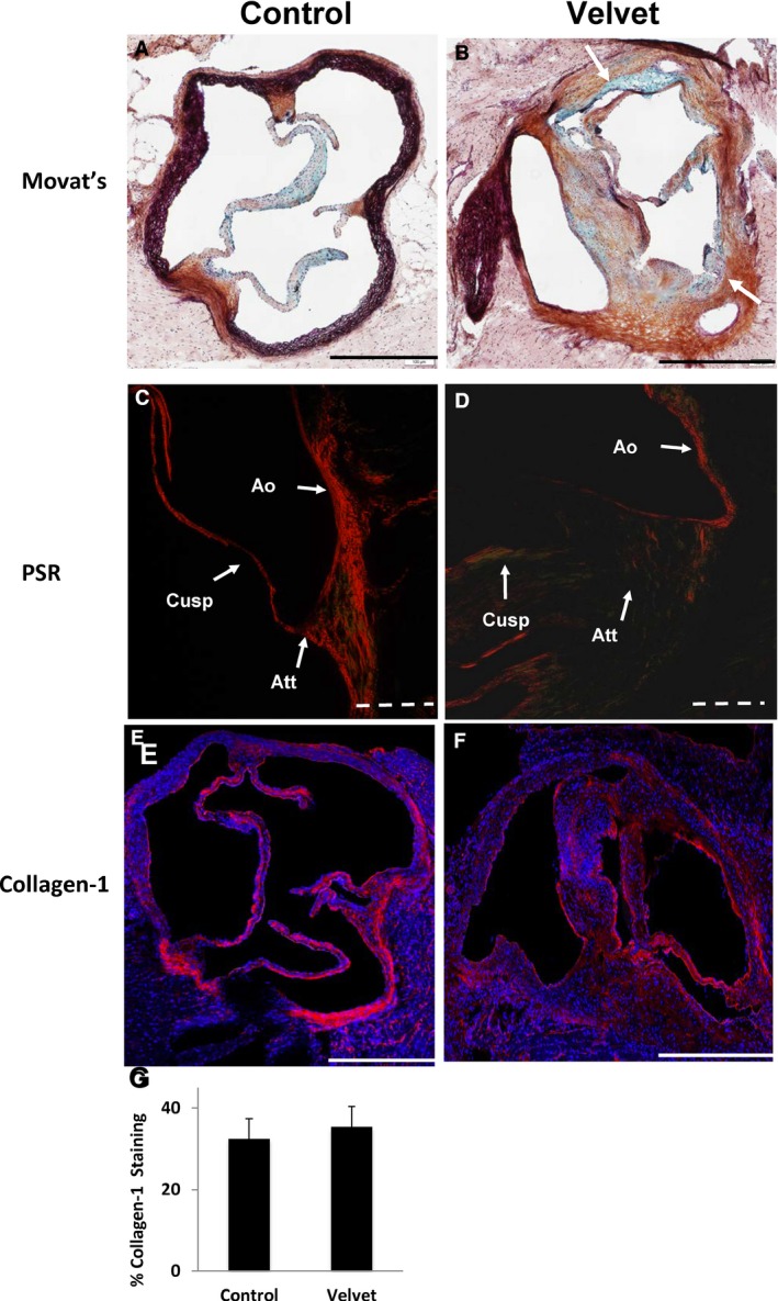
Extracellular matrix in aortic valves. A and B, Movat's Pentachrome staining shows discrete cusp attachment sites, which are composed mostly of collagen (brown), in the control valve. Control valve cusps contain mostly collagen and proteoglycan (blue). Demarcation between valve cusps and aortic wall is less distinct in the Velvet valve with both aortic regurgitation and aortic stenosis (white arrows). C and D, Picrosirius Red (PSR) staining viewed under polarized light depicts collagen fibers at site of cusp attachment (Att) oriented roughly parallel to the aortic wall (Ao) in the control valve. There is minimal transmission of polarized light, indicating lack of organization of collagen fibers, at the site of cusp attachment in the Velvet valve. E and F, Immunostaining for collagen‐1 (red) and cell nuclei (blue). G, Group data indicate similar proportion of valve tissue staining positive for collagen‐1 in Velvet vs control (N=6 each). Images and data were obtained from mice at 12 months of age. Solid bar=500 μm; dashed bar=100 μm. Data are given as mean±SEM.
