Figure 6.

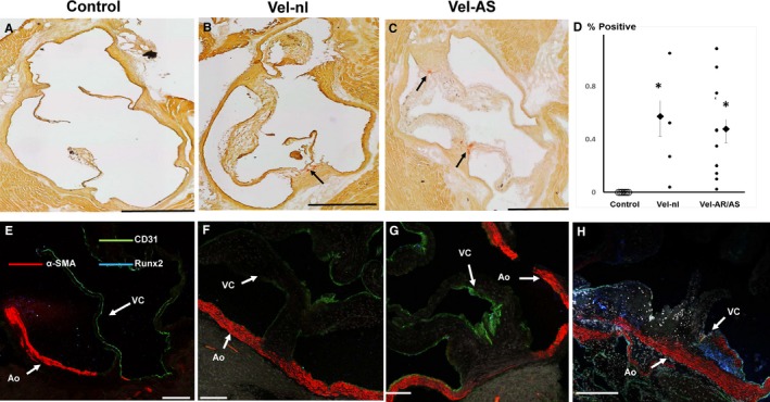
Valve interstitial cell transdifferentiation and calcification at 12 months of age. A through C, Alizarin Red staining shows modest calcification in Velvet valves (arrows) and none in control valve. D, Quantitation of Alizarin Red staining. Calcification was not greater in valves with echocardiographic evidence of moderate or severe aortic regurgitation (AR) or aortic stenosis (AS) than in Velvet valves with minimal valve dysfunction (Vel‐nl). Smaller points represent individual valves. Larger points and bars represent mean±SEM. E through G, Immunostaining for CD31, α‐smooth muscle actin (α‐SMA), and Runx2 in control and Velvet valves; α‐SMA is abundant in aortic wall (Ao), but not in valve cusps (VCs). Runx2 staining is minimal or absent. H, Immunostains from a Reversa valve, which is known to undergo valve interstitial cell transdifferentiation to myofibroblasts (α‐SMA) and osteogenic transdifferentiation (Runx2),16 serve as “positive control.” Black bar=500 μm. White bar=100 μm. *P<0.025 vs control.
