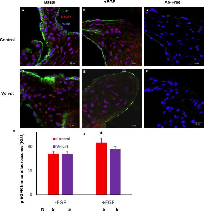
Figure 7.

Immunostaining for phosphorylated epithelial growth factor receptor (p‐EGFR) in the aortic valve. A and D, Under basal conditions, p‐EGFR (red) is present in both control and Velvet valves. B and E, After incubation with EGF, level of p‐EGFR may increase somewhat less in the Velvet valve than in the control valve. C and F, In the absence of anti‐CD31 and anti–p‐EGFR antibodies (Abs), there is negligible autofluorescence or secondary antibody fluorescence. G, Group data. Original magnification ×63. Bar=20 μm. RLU indicates relative light unit. *P<0.05 vs EGF.
