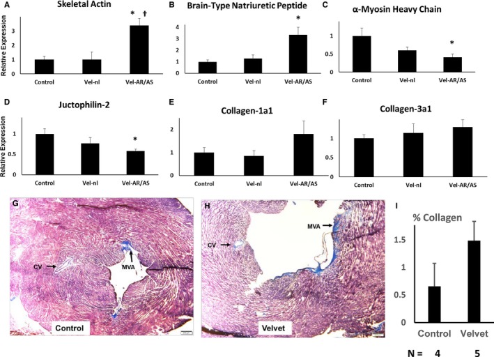
Figure 9.

Myocardial adaptation to hemodynamic stress at 12 months of age. A through F, Gene expression was first normalized to β‐actin expression, then normalized to expression in control myocardium. Results are pooled from 4 hearts from each group. G through I, Masson's Trichrome staining of left ventricular myocardium from a control mouse (left ventricular ejection fraction [LVEF], 0.79) and from a Velvet mouse (LVEF, 0.15). Staining in mitral valve annulus (MVA) served as positive control for collagen. Staining around coronary vessels (CVs) was seen occasionally in Velvet myocardium, but not in control myocardium. Small clear bar at lower right=200 μm. Vel‐nl indicates Velvet mice with normal or mildly impaired valve function; Vel‐AR/AS, Velvet mice with moderate/severe aortic regurgitation or aortic stenosis. *P<0.025 vs control; † P<0.05 vs Vel‐nl.
