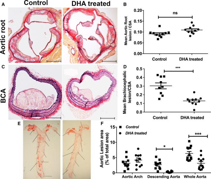
Figure 3.

Differential effects of DHA feeding on lesion development in different areas of the vascular beds. Male apoE−/− (apolipoprotein E–null) mice aged 8 weeks were fed a Western‐type HFD alone (control) or an HFD and jelly containing DHA (DHA treated; 300 mg/kg per day) daily over 12 weeks. A, Representative images of aortic roots stained with AB/EVG after 12 weeks of feeding (n=12 per group). Scale bars=100 μm. B, Mean lesion area of aortic root sections, normalized to CSA (n=12). Data are mean±SEM, analyzed by unpaired Student t test, P=ns. C, Representative images of brachiocephalic sections stained with AB/EVG (scale bars=100 μm). D, Mean lesion area of brachiocephalic arteries, normalized to CSA. Data are mean±SEM, analyzed by unpaired Student t test, 9 or 10 per group, ***P<0.001. E, Representative en face morphometric images of the total aortic lesion area and (F) whole aortic, aortic arch, and descending aortic lesion area calculated as a percentage of the total surface area of the whole aorta, showing significant reduction in the total lesion formation in the distal aorta in the DHA group compared with control. Data are mean±SEM, analyzed by 2‐way ANOVA and Tukey post‐test, 11 or 12 per group, *P<0.05, ***P<0.001. AB/EVG indicates alcian blue and elastic van Gieson; CSA, cross‐sectional area; DHA, docosahexaenoic acid (22:6n‐3); HFD, high‐fat diet.
