
Figure 6.
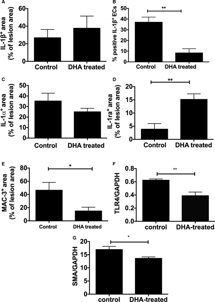
IL‐1 distribution in aortic atherosclerosis and lesion characteristics in response to DHA feeding. Male apoE−/− (apolipoprotein E–null) mice, from 8 weeks of age, were fed either an HFD alone or an HFD and DHA (300 mg/kg per day) daily. Following 12 weeks of diet, immunostaining, measured semiquantitatively as a percentage of total lesions, showed no difference in (A) IL‐1β or (B) IL‐1α. The number of IL‐1β–positive ECs, measured semiquantitatively as a percentage of total number of ECs, is lower in DHA‐treated mice compared with control (B). D, IL‐1ra is increased and (E) Mac‐3 (CD107b) is decreased in DHA‐treated animals. Image analysis was performed using NIS‐Elements software, and data are represented as mean±SEM, 6 to 8 per group. Student t tests indicate a significant difference with *P<0.05; **P<0.01. Western blot analysis of mouse aortas showed (F) TLR‐4 and (G) SMA levels are significantly decreased following DHA feeding. DHA indicates docosahexaenoic acid (22:6n‐3); EC, endothelial cell; HFD, high‐fat diet; IL, interleukin; SMA, smooth muscle actin; TLR‐4, Toll‐like receptor 4.