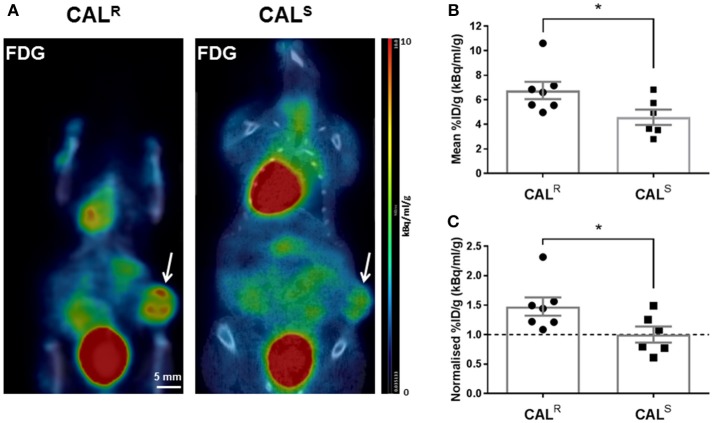
Figure 3.
(A) Coronal whole body PET-CT images of mice bearing CALR and CALS HNSCC xenografts (arrowed) acquired 1 h post injection of 18F-FDG. The images are shown on the same PET scale of 0–10% injected dose per gram (%ID/g). Marked uptake of 18F-FDG was also observed in the heart and bladder. (B,C) A significantly higher relative uptake of 18F-FDG was determined in the CALR cohort compared to the CALS tumors. Data are the individual (B) %ID/g and (C) normalized %ID/g for each tumor (relative to the average of the CALS cohort), and the cohort mean ± 1 s.e.m., *p < 0.05, Student's t-test.

